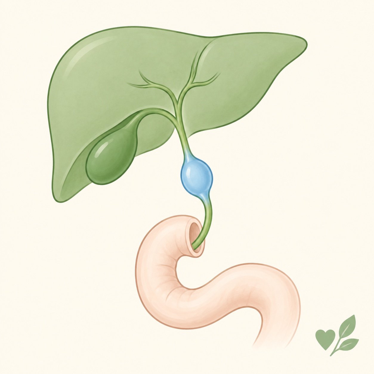
Most parents first hear the words “choledochal cyst” on a scan report. Your child may have had yellow eyes that come and go, bouts of tummy pain, or pale, chalky stools. Or a pregnancy scan may have shown a small fluid-filled bubble near your baby’s liver. Below is what the cyst is and why the whole cyst has to be removed rather than drained. It also explains why your child will need check-ups for life.
What Is a Choledochal Cyst?
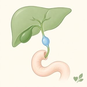
Bile is a digestive juice made in the liver. It travels to the intestine through a tube called the bile duct. In a choledochal cyst, part of this tube is ballooned out into a sac. Bile collects and moves slowly inside the sac. A child is born with this problem, although it may not cause symptoms until later. It is rare: about 1 in 13,500 in the United States and up to 1 in 1,000 in Japan. Girls are affected 3 to 4 times more often than boys.
Why It Happens
The pancreas is a gland that makes digestive juice. Its duct and the bile duct normally meet only a very short distance before they empty into the intestine. In most children with a choledochal cyst, the two ducts join too early, forming a long shared channel. This lets juice from the pancreas flow backwards into the bile duct. Over time, the juice irritates and weakens the duct wall, and the duct swells. Over many years, this irritation can turn the lining of the cyst abnormal. That abnormal lining can later become cancer. This is why the cyst is removed.
How It Shows Up in Babies and Older Children
The signs depend on age. In babies under one year, the usual signs are jaundice (yellow skin and eyes), pale or clay-coloured stools, and an enlarged liver. In older children, the problem often comes in episodes. There may be jaundice that comes and goes, tummy pain, or fever with infection of the bile duct (cholangitis). There may also be inflammation of the pancreas (pancreatitis). Many textbooks describe a “classic” trio of pain, jaundice and a lump in the upper right tummy. In reality, only about 20 out of 100 children have all three. So a child can have a cyst without any lump, or with only one of these signs. Some cysts are now first seen on a pregnancy or newborn ultrasound, before any symptoms at all.
How Doctors Confirm It
The first test is usually an ultrasound. It is simple and painless, and it picks up most cysts (between 71 and 97 out of 100). The best test to map the cyst is an MRCP, a special MRI scan of the bile duct and the duct from the pancreas. MRCP finds almost every cyst (90 to 100 out of 100) and uses no radiation. When I see a child with a choledochal cyst, I use the MRCP to see exactly where the two ducts join before planning the operation. Blood tests check the liver and pancreas. Some children also need vitamin K before surgery, because blocked bile flow can affect blood clotting.
Types of Choledochal Cyst
Doctors group choledochal cysts into five main types, depending on which part of the bile ducts is swollen. Type I is by far the most common, making up 90 to 95 out of 100 cysts. In Type I, the main bile duct outside the liver is ballooned. The other types are less common, and some involve the small bile ducts inside the liver as well. The type matters because it decides the exact operation and how closely the child needs to be watched afterwards.
Why the Whole Cyst Must Be Removed
Draining the cyst is not enough. It has to come out. In the past, some surgeons simply drained the cyst into the intestine. That approach has been abandoned. Draining leaves the damaged cyst lining behind, and that lining carries a risk of cancer later in life. It also allows repeated infections. For the common types (Types I and IV), the standard treatment is to remove the whole swollen bile duct outside the liver. In Type IV, some cysts lie inside the liver and cannot always be removed, which is why follow-up matters even more. The surgeon then joins the healthy bile duct to a loop of the child’s own small intestine, so that bile drains freely again. Doctors call this new bile route a Roux-en-Y hepaticojejunostomy. When the surgeon has experience with this operation, it is increasingly done by keyhole (laparoscopic) surgery. Rarer types are treated differently. Some need only a smaller operation, and some are treated through an endoscope (a thin camera tube). A very rare type inside the liver may need part of the liver removed.
When Should the Operation Be Done?
Surgeons do not leave these cysts alone. If the cyst was found on a pregnancy scan and your baby is well, some surgeons operate in the first few months. Others plan the operation within the first year of life. Both aim to protect the liver from slow scarring. They also aim to prevent infection of the bile duct or a burst cyst. If your child already has symptoms, surgeons operate early rather than wait. Please see a pediatric surgeon promptly.
Life After Surgery
Most children do well after the operation. The problems below can still appear years later. The most common late problem is a narrowing (stricture) at the join between the bile duct and the intestine. A narrowing can slow bile flow and lead to stones inside the liver ducts. These stones can appear anywhere from 3 to 22 years after surgery. Some children also have repeated pancreatitis. Removing the cyst greatly lowers the cancer risk, but does not bring it to zero. This is especially true in types where cysts also involve the ducts inside the liver. For these reasons, follow-up must continue for life, with regular check-ups and scans, even when the child feels completely well.
What Every Parent Must Know
- Yellow eyes, pale stools or tummy pain can all point to a cyst. Do not wait for all three signs together.
- The cyst must be removed, not drained, because of the cancer risk.
- A well baby with a cyst seen on a scan still needs an operation, in the first months or within the first year.
- A child with symptoms should see a pediatric surgeon promptly.
- Problems can appear many years after surgery, so check-ups are for life.
When to See a Doctor
See a doctor urgently if your child has yellow eyes or skin, pale or white stools, or tummy pain with fever. Did a pregnancy or newborn scan show a cyst near the liver? Then arrange an early visit to a pediatric surgeon, even if your baby looks healthy. A child who has already had the operation should never miss follow-up. That child should also be seen quickly if jaundice, fever or tummy pain returns.
Dr. Tanmay Motiwala is a pediatric surgeon in Raipur, Chhattisgarh, trained at AIIMS Jodhpur. He assesses and treats children with choledochal cysts and other bile duct and liver problems from across Chhattisgarh and central India, including complete removal of the cyst and long-term follow-up.
Related reading:
- My Newborn Is Still Yellow After Two Weeks — When Is Jaundice Dangerous?
- Appendicitis in Children: When Stomach Pain Needs Surgery
- The Scan Shows My Baby’s Kidney Is Swollen — Should I Worry?
📋 This article is part of Dr. Motiwala’s Pediatric General & Abdominal Surgery in Raipur services — see the full range of conditions treated, what to expect, and when to see a pediatric surgeon.
Related conditions parents also read
- Newborn still yellow after two weeks (biliary atresia)
- When stomach pain needs surgery (appendicitis)
- Something seen on the pregnancy scan (swollen baby kidney)
Worried about your child? Dr. Tanmay Motiwala consults in Raipur, Jagdalpur & Rajim. Book an appointment or call +91 83190 84711.
⚠️ Important Disclaimer: This article is for general information and educational purposes only. It is not a substitute for professional medical advice, diagnosis, or treatment. Every child’s condition is different — facts, prognosis, and management can vary significantly from case to case. Please consult a qualified pediatric surgeon for advice specific to your child.
Sources: Coran’s Pediatric Surgery (7th ed), Ch. 106 (Choledochal Cyst), Gonzales & Lee; Rob & Smith’s Operative Pediatric Surgery (7th ed).